|
Laboratory Materials |
|
Return to Table of Contents
Instructions Topic 8. Fast and slow motor axons.
________________ (Name) I. MATERIALS. Live locusts or grasshoppers, the larger, the better (nymphs are okay). Nerve stimulator, Grass SD-9, or equivalent. Wire stimulating electrodes, or grease stimulating electrodes. Audio amplifier and speaker. A fresh tube of Vaseline (older Vaseline yellows and is toxic to the nerves). A micromanipulator for the electrodes. Dissecting microscope. Insect saline. A video camera capable of projecting the grasshopper image onto a screen.
Prepare the dissection before the class, if possible. Allow time for one failure. Be sure to practice the preparation. The dissection can be attempted by advanced undergraduates with an aptitude for micromanupulation and depending on available equipment. Alternatively, the class can learn as much by watching the TA stimulate the slow and fast nerves, or taking over the stimulator once the electrodes are in place.
II. LEARNING OBJECTIVES. Each student should be able to: 1. Define slow and fast motor axons and the responses they evoke. 2. Understand the jump mechanism of the extensor tibia muscle.
How: Stimulate the nerves supplying the extensor tibia muscle and identify the exact muscles responsible for the resulting movements. Identify the response to stimulation of the fast extensor and flexor tibia nerves by the movements of the tibia. Observe actions of slow axon stimulation on the extensor tibia muscle. Identify Heitler's hump near the femur-tibia joint.
III. INTRODUCTION. Insect muscles are composed of one or more muscle fibers (a fiber is a single muscle cell). All insect muscles are multiterminally and usually polyneuronally innervated. Polyneuronal means that more than one type of motor neuron supplies each muscle fiber of a given muscle. Multiterminal innervation means a single motor axon branches to supply nerve endings over the entire surface of an individual muscle cell.
Insect muscles can be innervated by as many as 12 motor neurons. However, other muscles are innervated by only one, or just a few, motor neurons. The fibrillar flight muscle fibers of the cyclorrhaphan Diptera are all innervated by a single excitatory motor neuron. The object of this demonstration, the extensor tibia muscle of the hind leg, or jumping leg of the grasshopper, is an extremely large muscle, one of the largest leg muscles in insects. It can generate 800 grams of force in a large adult locust in producing a normal kick response, but can also be finely positioned. Remarkably, all of these movements are controlled by only three motor neurons supplying each leg. The motor neurons are named the fast, slow and inhibitory motor units.
The slow motor neurons are defined by the repines of the muscle to stimulation of the axon. Slow axons produce no response when shocked singly, but graded movements result when the stimulation frequencies are high, above about 15 shocks/second in the case of the extensor tibia muscle. The higher the frequency of stimulation, the greater the tension developed in the muscle, until maximum flexion is obtained near the limits of the stimulation, somewhere around 200 shocks/second. By varying the frequency of stimulation of a single slow axon, one is able to adjust the limb attached to the muscle apodeme to any position within the full flexed or extended travel of the limb.
The slow units are designed to be able to sustain muscle contraction for long periods of constant use. This means there are numerous very large terminals, that are full of mitochondria positioned to supply energy to support sustained operation for long periods of frequent use. The associated synaptic synthesis machinery is also geared to generate and recycle large amounts of neurotransmitter chemical.
The fast units are at the opposite end of the spectrum. Fast motor axons are designed to generate immediate responses, and not to sustain activity for long periods. Thus the fast units fatigue rapidly, failing a short time after being placed under sustained stimulation. This means their cellular machinery is geared to release large amounts of neurotransmitter in response to single stimuli which produce maximal responses in the postsynaptic muscle receptors. There are relatively few mitochondria in the nerve terminals because neurotransmitter is not designed to be supplied constantly in high volume under sustained work load conditions.
Muscle fibers themselves reflect these differences in function with tonic fibers being full of mitochondria with the contractile filaments being long, while phasic muscle fibers have relatively few mitochondria with short myofibrils and a very well-developed sarcoplasmic reticulum.
The extensor tibia muscle is unique among insects in having the slow and fast axons traveling to the same muscle my different nerve bundles. This quirk of the anatomy allows the fast extensor tibia axon to be stimulated by itself, then the slow extensor tibia axon to be stimulated separately. If both units were in the same nerve bundle, it would be difficult to stimulate one of them alone.
IV. DIRECTIONS. Because the preparation of the slow and fast motor units is involved and requires practice to perform on call, it will be prepared by the TA.
A fresh grasshopper is pithed, then mounted dorsum down in a wax dish. Splay the legs out at a 45 degree angle from the body and place crossed pins over the distal end of the femur to hold the femur in place. Be very careful exert too much pressure on the coxal, or the leg will be aborted and the connection to the body broken.
Once the body is secured, cut away the bottom of the thorax. This will be difficult because the presence of strong sutures for internal flight muscle attachments makes the cuticle here particularly hard and difficult to remove. A scalpel may be needed. The metathoracic ganglion should be exposed enough to identify the ventral nerve cord connectives running into abdomen, and the very large crural nerve running into the hind legs. If these features are not clearly visible, dissect more until the entire metathoracic ganglion is exposed.
Moisten with a few drops of saline and apply stimulation electrodes to the crural nerve. The crural nerve is larger that the connectives of the ventral nerve cord at this level of the body and is conspicuously larger than any other nerves leaving the ganglion. It should be seen projecting into the femur. Set the stimulator for multiple shocks and the frequency for about one per second. Turn the voltage to minimum and turn on the stimulator. Advance the voltage until twitches are seen in the leg.
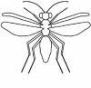
Because the crural nerve has many axons besides the fast extensor tibia unit, when the voltage is increased, that unit will respond with the lowest threshold for response, not necessarily the extensor tibia fast axon. Four blank spaces are provided below to identify the direction of twitch and name the muscle stimulated. Identify the direction, then list the name of the muscle responsible. A diagram of the locust leg is provided for reference.
Direction of twitch Muscle responsible
1. ____________________________ _________________________
2. ____________________________ _________________________
3. ____________________________ _________________________
4. ____________________________ _________________________
Figure of the locust leg with leg muscles named.
Figures 106-9, pp. 163-169, muscles of the metathoracic appendages. In: J. C. Jones, The Anatomy of the Grasshopper, Charles C. Thomas, Springfield, IL, 1981, or the equivalent.
V. SUMMARY ANALYSIS. What was learned in this exercise?
VI. REFERENCES Hoyle G. 1968. Slow and fast axon control of contraction in insects. In: Experiment in Physiology and Biochemistry, ed by G. A. Kerkut, Academic Press, London, pp. 287-293.
Jones, Jack C. 1981. The Anatomy of the Grasshopper (Romalea microptera), Charles C. Thomas, Springfield, Illinois.
Miller, T. A. 1979. Insect Neurophysiological Techniques, Springer-Verlag, New York.
_________________ (Name) |
 |
Click on the picture to go to Dr. Miller's Lab Web Page. |

|
More Topics on the Wing |
 |
|
About Us |
 |
Click on Picture to go to the link |
Page Designed by
Harald Baella. Last updated
01-25-05
Copyright © 2003-05 Miller Web Design.