|
Laboratory Materials |
|
Return to Table of Contents
Instructions Topic 2. Insect endocrine system ________________ (Name) I. MATERIALS. Manduca sexta larvae, last instar was too large for this exercise. Use an orthopteroid insect such as a cricket or cockroach. Last instar housefly, Musca domestica, or some other cyclorrhaphan Dipteran of moderate size, but not a Drosophila species (too small). Waxed Dental floss for tying body ligations. Dissecting scissors, Paper towels, Petri dishes. Eclosion-Triggering hormone, Injection needle.
II. LEARNING OBJECTIVES Each student should be able to identify the main endocrine glands in insects to help understand growth and development and the source of insect hormones.
How: Observation of major endocrine glands in an experimental insect. You will dissect insect brains containing a variety of neurosecretory cells. Perform the famous "ligation experiment," one of the simplest and yet most powerful tools in insect physiology showing the functions of insect hormones.
III. INTRODUCTION. One of the original research questions in insect physiology was "Do insects use hormones to control their growth and development?" The answer to this question, an emphatic Yes, was demonstrated by the Polish physiologist, Stefan Kopec using extremely simple experimental methods.
In 1917 Stefan Kopec first demonstrated a "head critical period" in insects by ligating larvae of the gypsy moth, Lymantria dispar. He showed that either both halves of the larvae pupated, or only the anterior end pupated, leaving a permanent larval posterior. The difference in these experimental animals was the time at which the ligation was performed. If the last instar larvae were tied too short a time before the next molt, the entire animal pupated, but when ligated a longer time before the molt, only the front end pupated. The time defined by these results became know as the head critical period.
Kopec further demonstrated that the brain was responsible for pupation. Animals debrained before the head critical period formed permanent larvae. Brains inserted into permanent larvae induced pupation and hemolymph from larvae obtained after the head critical stage also induced pupation in permanent larvae. These results established that a blood-borne factor (or hormone) associated with the brain was responsible for molting (or pupation) in insects.
Kopec's experiments were overlooked at first, but they were rediscovered around 1934 and popularized by Sir Vincent Wigglesworth and others who pointed out their significance, and showed that they could be repeated on a variety of insects, both hemi- and holometabola.
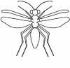
IV. DIRECTIONS. 1. Observation of insect endocrine glands. The brain, corpora cardiaca, corpora allata and prothoracic glands will be observed in Periplaneta americana. The location of cells in the brain that synthesize prothoracicotropic hormone will be demonstrated from publications (see references below).
The corpora cardiac and allata are connected to the posterior part of the brain and usually form a combined tissue, called the retrocerebral complex. The prothoracic gland is generally located in the thorax and consists of loosely connected strands of very large cells. The TA will demonstrate how to dissect and identify these organs. Label the figure below with the proper name based on your preparation, that of a colleague, or the TA. And draw the chemical structure of beta-ecdysone (page 405, Fig 13.6 in Gillott) with an arrow showing where it is biosynthesized.
2. Demonstration of head critical period (HCP). Insects grow by a series of molts. They generally expand the surface of the epidermis under the old cuticle in preparation for a molt and unfold the new cuticle and therefore increase in size immediately after a molt. To initiate molting, the brain must be present before the head critical period (HCP).
For Manduca sexta (This exercise is optional and should only be attempted by someone experienced with the tobacco hornworm.):
1) prepare 3 groups (3 larvae each) of last instar larvae. (i) group 1; 3 larvae within 24 hrs after molting which are before or during the HCP. (ii) group 2; 3 larvae 4 days after molting which are after the HCP (iii) group 3; larvae within 24 hrs after molting which are before or during the HCP.
2) Ligate all larvae between the first and the second abdominal segments with thin string (dental floss). Prepare two loops of a simple granny knot. Position the loop on the body. Pull the ends of the string and be careful not to pull the ends to tightly. Tie the loose ends in another single granny knot and tighten this too. Snip off the loose ends of the string and return the ligated larvae to the holding container.
Full page drawing of cockroach in outline with label lines for:
Brain.
Corpora cardiaca.
Corpora allata.
Prothoracic gland.
Heart.
Ventral nerve cord.
3) Inject Eclosion-Triggering Hormone into the posterior part of group 3 larvae.
4) Place groups 1, 2 and 3 in plastic Petri dishes with diet and wet paper towel and keep in an incubator.
5) Results will be observed and interpreted during the next demonstration session in one week.
For housefly, Musca domestica: 1) Obtain young third instar larvae. Ligate ten larvae in the middle as described above. It may help to cool the larvae by placing them on ice for a short time before attempting the ligation. 2) Repeat step one with another ten old larvae immediately before pupation. Old larvae often clear the gut when preparing for pupation. Younger larvae have a conspicuous dark line that can be seen inside in the middle of the body that is food material being digested. Older larvae lack this dark line. 3) Repeat this procedure (place ten larvae on ice in the same manner as the others) but do not ligate. These will serve as sham controls. Return to culture. 4) Return all larvae to their rearing media and place in storage. Analyze at each succession class meeting by counting the number that have pupated and indicate the appearance of the pupated individuals. Use drawing.
In the spaces below indicate the appearance of the ligated insects after one week. [TA: ligated insects from all students can be pooled instead of kept separate.]
1. Sham control insects. 2. Ligated old larvae. 1. 1. 2. 2. 3. 3. 4. 4. 5. 5. 6. 6. 7. 7. 8. 8. 9. 9. 10. 10.
3. Ligated young larvae. 1. 2. 3. 4. 5. 6. 7. 8. 9. 10.
V. SUMMARY ANALYSIS. Describe in your own words what you have learned from this exercise.
VI. REFERENCES.
Chen, D. H. 1968. American cockroach. In: Experiments in Physiology and Biochemistry, ed by G. A. Kerkut, Vol. I, pp. 201-208.
Gillott, C. 1995. Entomology, 2nd edn., Plenum Press, New York, NY pages: 402-406.
Guthrie, D. M, and A. R. Tindall. 1968. The Biology of the Cockroach, Edward Arnold, London, see page 159, diagram of cockroach brain.
Nijhout, H.F. 1994. Insect Hormones, Princeton University Press, Princeton, NJ.
Zitnan, D. T.G. Kingan, S.J. Kramer and N.E. Beckage. 1995. Accumulation of Neuropeptides in the Cerebral Neurosecretory System of Manduca sexta Larvae Parasitized by the Braconid Wasp Cotesia congregata. J. Comp. Neurol. 356: 83-100. _________________ (Name) |
 |
Click on the picture to go to Dr. Miller's Lab Web Page. |
|
More Topics on the Wing |
 |
|
About Us |
 |
Click on Picture to go to the link |
Page Designed by
Harald Baella. Last updated
01-25-05
Copyright © 2003-05 Miller Web Design.